Home
/ Diagram Of Animal Cell Membrane / Membranes And Membrane Lipids - Animal cell functions are solely dependent on the organelles and structures associated with the cell.
Diagram Of Animal Cell Membrane / Membranes And Membrane Lipids - Animal cell functions are solely dependent on the organelles and structures associated with the cell.
Diagram Of Animal Cell Membrane / Membranes And Membrane Lipids - Animal cell functions are solely dependent on the organelles and structures associated with the cell.. The contents of a cell are called the protoplasm. Animal cell membrane diagram page for you to see. The membranes lipid bilayer is mainly 2 layers of phospholipids; Draw a table of differences between the two cell types in the space provided. The membrane has 2 layers of phospholipids (fats with phosphorous attached), which at body temperature are like vegetable oil (fluid).
This animal cell diagram doesn't represent any particular animal cell, it provides insight into the primary organelles and the internal structure of the most animal cell. Diagram of plasma membrane (cell membrane), created with biorender.com. In addition to the phospholipid bilayer, the cell membrane also one important sterol is cholesterol, which regulates the fluidity of the cell membrane in animal cells. The diagram, like the one above, will include labels of the major parts of an animal cell including the cell membrane, nucleus, ribosomes, mitochondria, vesicles, and cytosol. These are organelles pertinent to plant cells.
Draw A Labelled Diagram Of Plant Cell And Animal Cell Brainly In from hi-static.z-dn.net Cholesterol molecules are selectively dispersed between membrane phospholipids. The membrane has 2 layers of phospholipids (fats with phosphorous attached), which at body temperature are like vegetable oil (fluid). In fact, the collective work of the. Removing cellular waste products from the cell. The contents of a cell are called the protoplasm. It is the outermost part of the cell in animals. Destroying the organelles that are not functioning properly. Animal cells are generally smaller than plant cells and lack a cell wall and chloroplasts;
Animal cell functions are solely dependent on the organelles and structures associated with the cell.
Animal cells, plant cells, prokaryotic cells, and fungal cells have plasma membranes. Animal, plant, protist, and fungus cells. It is the outermost part of the cell in animals. An animal cell diagram is a great way to learn and understand the many functions of an animal cell. Essentially, a cell membrane is the outermost barrier that separates the internal contents of a cell in the cytoplasm from the external environment (e.g. Note that they are composed of phospholipid molecules and protein. The cell membrane is the boundary that separates the inside of the cell from the outside of the cell. The cells of animals are. The diagram, like the one above, will include labels of the major parts of an animal cell including the cell membrane, nucleus, ribosomes, mitochondria, vesicles, and cytosol. Diagram of plasma membrane (cell membrane), created with biorender.com. Drawing of the fluid mosaic model. Start studying animal cell diagram. How do you tell eukaryotic cells from prokaryotic cells?
This animal cell diagram doesn't represent any particular animal cell, it provides insight into the primary organelles and the internal structure of the most animal cell. Animal, plant, protist, and fungus cells. The contents of a cell are called the protoplasm. Animal cells, plant cells, prokaryotic cells, and fungal cells have plasma membranes. Think of the cell membrane like the border control of the cell, controlling what comes in and what goes out.
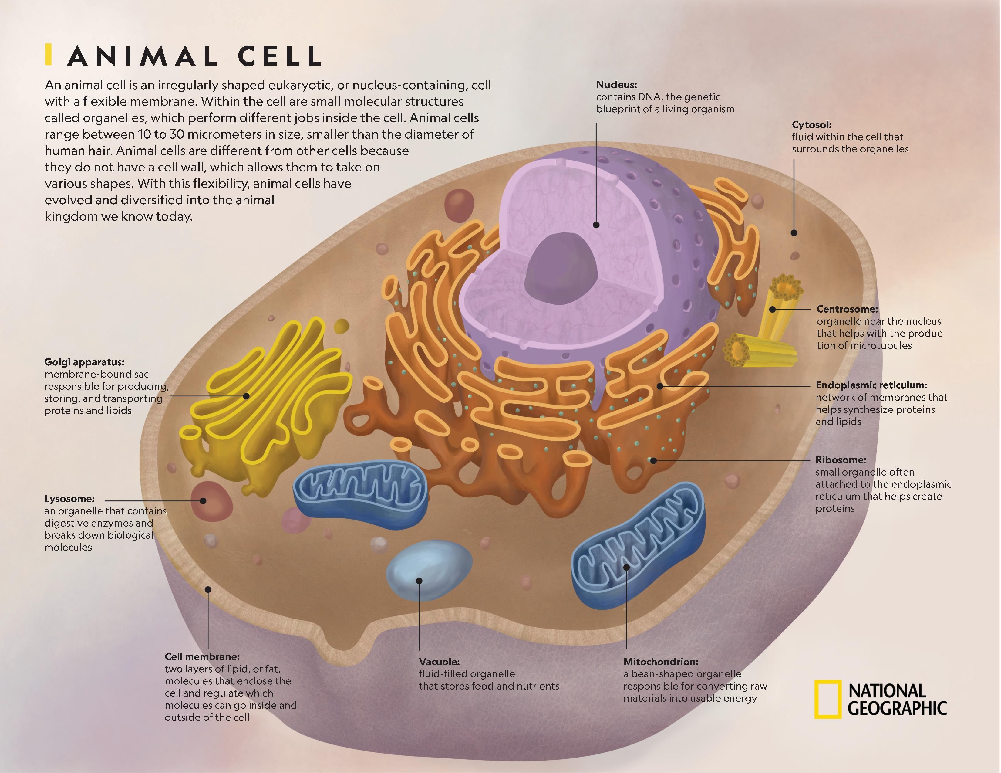
Animal Cell National Geographic Society from media.nationalgeographic.org This animal cell diagram doesn't represent any particular animal cell, it provides insight into the primary organelles and the internal structure of the most animal cell. Smooth endoplasmic reticulum, mitochondria, golgi bodies, lysosomes. These are organelles pertinent to plant cells. Start studying label cell membrane. Cells are covered by a cell membrane and come in many different shapes. While the animal cell functions and organelles are linked to each other. Cell membranes, also called the plasma membrane, is a physical barrier between a cell and the surrounding environment. The membranes lipid bilayer is mainly 2 layers of phospholipids;
The contents of a cell are called the protoplasm.
Drawing of the fluid mosaic model. Cholesterol is a component of animal cell membranes. All cells have a cell membrane around them. Start studying animal cell diagram. Unlike the eukaryotic cells of plants and fungi, animal cells do not have a cell wall. In truth, there are still features of plant and animal cells we're only lately discovering. Destroying the organelles that are not functioning properly. It is the outermost part of the cell in animals. The role and function of the plasma membrane; Structure of membrane in prokaryotes cell: Outermost in animal cell and lies next to cell wall in plant cell. All animal cells contain organelles. Animal, plant, protist, and fungus cells.
Animal cells, plant cells, prokaryotic cells, and fungal cells have plasma membranes. Essentially, a cell membrane is the outermost barrier that separates the internal contents of a cell in the cytoplasm from the external environment (e.g. In truth, there are still features of plant and animal cells we're only lately discovering. All animal cells contain organelles. The membranes lipid bilayer is mainly 2 layers of phospholipids;
Sgenofamthe Animal Cells Diagram from www.classroomjr.com That cells can be of different shapes and sizes. Unlike the eukaryotic cells of plants and fungi, animal cells do not have a cell wall. Essentially, a cell membrane is the outermost barrier that separates the internal contents of a cell in the cytoplasm from the external environment (e.g. The animal cell is made up of several structural organelles enclosed in the plasma membrane, that enable it to function properly, eliciting mechanisms that benefit the host (animal). Start studying label cell membrane. Cholesterol in mammalian membranes reduces membrane fluidity and permeability to some solutes. The ones mentioned on this page include centrosomes, goli apparatus, lysosomes, mitochondria, the nucleus and its parts such as the nuclear membrane and nuclear pores, also other. Diagram of plasma membrane (cell membrane), created with biorender.com.
Animal, plant, protist, and fungus cells.
Animal cells, plant cells, prokaryotic cells, and fungal cells have plasma membranes. The ones mentioned on this page include centrosomes, goli apparatus, lysosomes, mitochondria, the nucleus and its parts such as the nuclear membrane and nuclear pores, also other. Study the two diagrams of plant and animal cells below. Animal cell membrane diagram page for you to see. While the animal cell functions and organelles are linked to each other. These are organelles pertinent to plant cells. Drawing of the fluid mosaic model. All animal cells contain organelles. Essentially, a cell membrane is the outermost barrier that separates the internal contents of a cell in the cytoplasm from the external environment (e.g. This organelle is also referred to as plasma membrane. Animal cells are generally smaller than plant cells and lack a cell wall and chloroplasts; The cell membrane is the boundary that separates the inside of the cell from the outside of the cell. The cell membrane (also known as the plasma membrane (pm) or cytoplasmic membrane, and historically referred to as the plasmalemma) is a biological membrane that separates the interior of all cells from the outside environment (the extracellular space).
Share :
Post a Comment
for "Diagram Of Animal Cell Membrane / Membranes And Membrane Lipids - Animal cell functions are solely dependent on the organelles and structures associated with the cell."


Post a Comment for "Diagram Of Animal Cell Membrane / Membranes And Membrane Lipids - Animal cell functions are solely dependent on the organelles and structures associated with the cell."