Home
/ Drawing Structure Of Animal Cell Diagram : Draw A Well Labeled Diagram Of Animal Cell And Plant Cell Mention Major Function Of Each Organelle Brainly In - Here is a summary of their structure and function.
Drawing Structure Of Animal Cell Diagram : Draw A Well Labeled Diagram Of Animal Cell And Plant Cell Mention Major Function Of Each Organelle Brainly In - Here is a summary of their structure and function.
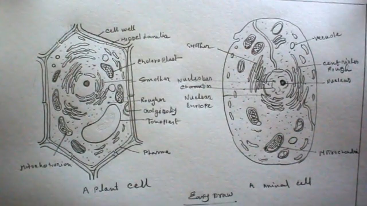
Drawing Structure Of Animal Cell Diagram : Draw A Well Labeled Diagram Of Animal Cell And Plant Cell Mention Major Function Of Each Organelle Brainly In - Here is a summary of their structure and function.. These are both specific types of cells, and the diagram is very clear, and labeled; Every animal cell has two of these small organelles (made of microtubules) and they help organize cell division (like a teaching assistant who help out near the office). Vacuoles in animal cells are many and small. Since animal cells lack a rigid cell wall it allows them to develop a great diversity of cell types, tissues, and organs. Plant cell and animal cell fall under eukaryotic type.
In truth, there are still features of plant and animal cells we're only lately. Plant cell are more consistent in size and shape and are surrounded by rigid cell wall, made up of cellulose. Plant cell and animal cell fall under eukaryotic type. Cell biology (biomedical laboratory science students). Internal structure of human heart shows four chambers viz.
How To Draw Plant Cell And Animal Cell Plant Cell Drawing Animal Cell Drawing Plant And Animal Cell Youtube from i.ytimg.com Vacuoles in animal cells are many and small. In fact, the collective work of the animal cell parts is responsible for overall functioning. It contains a number of structures known as organelles of. The nerves and muscles are made up of specialized cells that plant. Animal cell anatomy diagram structure with all parts nucleus smo. 2.3.2 annotate the diagram from 2.3.1 with the functions of each named structure. 720 x 1280 pixel type jpg download. In the labeled animal cell diagram animal cell functions and organelles are linked to each other.
This will also help you to draw the structure and diagram of an animal cell.
In the labeled animal cell diagram animal cell functions and organelles are linked to each other. Rough er is rough because it has ribosomes attached to its surface and ribosomes are cell structure that make protein (like the cooks. Animal and plant cell energy cycle vector illustration diagram with mitochondrion and chloroplast. It contains a number of structures known as organelles of. I spelt it wrong in the diagram, sorry). Animal cells have a single highly complex and prominent golgi apparatus. How to draw an animal cell diagram homework help doodledrawart image information: The structure of the plasma membrane. Lets us discuss the animal cell, types of an animal cell, animal cell diagram, its structure. Let`s draw a typical animal cell. In almost every video all of these membranes that we draw either the outer cellular membrane or the membranes of these organelles these are all lipid bilayers or phospholipid. The structure of an animal cell differs slightly from a plant cell, in terms of shape, protective covering and organelles. Prokaryotic cells (above) are much simpler in structure than eukaryotic cells.
Animal cells differ from plant cells in several regards though, including the lack of vacuoles if you wish to draw a particular cell such as an amoeba or paramecium, study them first. The largest organelle within the cell. 720 x 1280 pixel type jpg download. Every animal cell has two of these small organelles (made of microtubules) and they help organize cell division (like a teaching assistant who help out near the office). Drawing, graphic, biology animal cell anatomy diagram structure with all parts nucleus smooth rough endoplasmic reticulum cytoplasm golgi apparatus.
How To Draw Plant Cell And Animal Cell Step By Step Very Easy Youtube from i.ytimg.com Animal cell structures, functions & diagrams. Check this diagram and learn m. Growth of reproduction the protoplasm inside the cell membrane and outside the nucleus is known as cytoplasm. 2.3.1 draw and label a diagram of the ultrastructure of a liver cell as an example of an animal cell. Animal cells differ from plant cells in several regards though, including the lack of vacuoles if you wish to draw a particular cell such as an amoeba or paramecium, study them first. A system of flattened membranes called cisternae (mainpoint: The cell (from latin cella, meaning small room) is the basic structural, functional, and biological unit of all known organisms. Though this animal cell diagram is not representative of any one particular type of cell it provides insight into the primary organelles and the intricate internal structure of most animal cells.
I based my information on this diagrams
The animal cell is more fluid or elastic or malleable in structure; Cells are the smallest units of life. Bacteria and the parasite that causes malaria consist of single cells, while plants and animals are made up of a drawing of the cell as seen with an electron microscope is shown in diagram 3.3. Every animal cell has two of these small organelles (made of microtubules) and they help organize cell division (like a teaching assistant who help out near the office). Plant cell are more consistent in size and shape and are surrounded by rigid cell wall, made up of cellulose. But at the same time it is interpretive. The plant cell as more rigid and stiff walls. It is also called the cytosome. Structure of animal cell and plant cell under microscope diagrams image information: They are eukaryotic cells, that means they contain a membrane bound nucleus. Cellular water levels biological vector illustration diagram with animal and plant cell. This is a diagram of an animal cell. Extracellular structures and intercellular junctions.
Here is a summary of their structure and function. This is a diagram of an animal cell. I based my information on this diagrams Rough er is rough because it has ribosomes attached to its surface and ribosomes are cell structure that make protein (like the cooks. Growth of reproduction the protoplasm inside the cell membrane and outside the nucleus is known as cytoplasm.
Human Animal Cell Structure Animal Human Cell Parts Diagram Diagram With Computer Components Clean White Sheet Without Explanation No Description Education Science Biology Image 2d Drawing Stock Vector Adobe Stock from as2.ftcdn.net Animal cells have an irregular shape and structure and bend and fold easily. That's the major difference between plant and animal cells under microscope. Animal cell anatomy diagram structure with all parts nucleus smo. Two atria and two ventricles and couple of blood vessels opening into them. The size of the animal cell ranges from a few millimetres to microscopic micron. The extracellular matrix and cell wall. Structure of a typical animal cell. Unlike the eukaryotic cells of plants and fungi, animal cells do not have a cell wall.
You know, animal cell structure contains only 11 parts out of the 13 parts you saw in the plant cell diagram, because chloroplast and cell wall are available only in a plant cell.
In almost every video all of these membranes that we draw either the outer cellular membrane or the membranes of these organelles these are all lipid bilayers or phospholipid. But at the same time it is interpretive. Two atria and two ventricles and couple of blood vessels opening into them. Generalized cell is used for structure. It contains a number of structures known as organelles of. Animal cells differ from plant cells in several regards though, including the lack of vacuoles if you wish to draw a particular cell such as an amoeba or paramecium, study them first. Plant cell and animal cell fall under eukaryotic type. There are usually some other structures like flagella, cilia. I spelt it wrong in the diagram, sorry). Rough er is rough because it has ribosomes attached to its surface and ribosomes are cell structure that make protein (like the cooks. Animal cells have a single highly complex and prominent golgi apparatus. In truth, there are still features of plant and animal cells we're only lately. These are both specific types of cells, and the diagram is very clear, and labeled;
Share :
Post a Comment
for "Drawing Structure Of Animal Cell Diagram : Draw A Well Labeled Diagram Of Animal Cell And Plant Cell Mention Major Function Of Each Organelle Brainly In - Here is a summary of their structure and function."


Post a Comment for "Drawing Structure Of Animal Cell Diagram : Draw A Well Labeled Diagram Of Animal Cell And Plant Cell Mention Major Function Of Each Organelle Brainly In - Here is a summary of their structure and function."